Analyze skin lesions to help detect early signs of skin cancer

Thomas Oldenburger

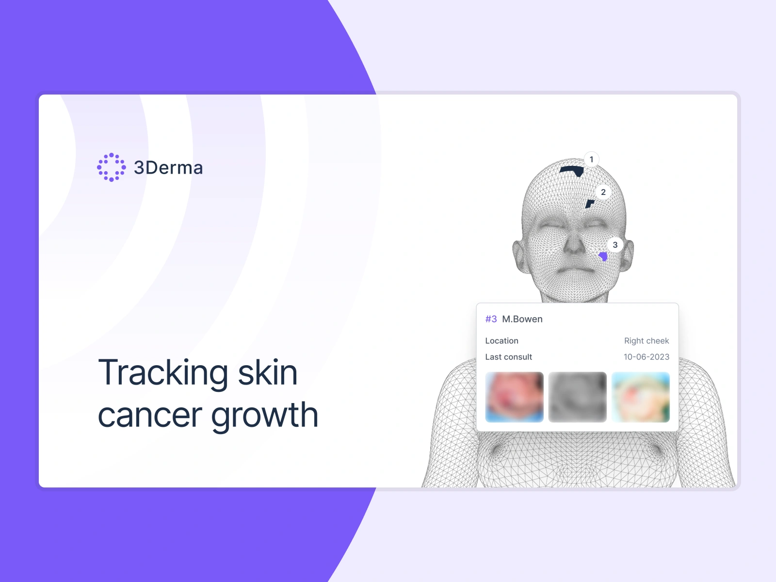
Skin cancer is tracked on paper diagrams.
We fixed that.
A skin cancer patient sees a dozen providers over a decade. We designed 3Derma so each of them opens the file and sees the same 3D map: every lesion, in its actual place, with its whole history attached.
The handoff is where tracking breaks
A skin cancer patient is rarely seen by one person. Over the years the file passes between dermatologists, surgeons, residents, and back again. Each provider inherits the history: notes in the EHR, photos in a folder, locations sketched on a paper body diagram.
The doctor who treated a lesion in 2016 usually isn't the one looking at it in 2023. So the first minutes of every consult go to reconstructing which spot was which, and what was done to it.
3Derma fixes each lesion to a point on a 3D model of the patient's body and attaches the whole history to that point. Any provider who opens the file sees the same map. We designed the product around that idea.
What we did
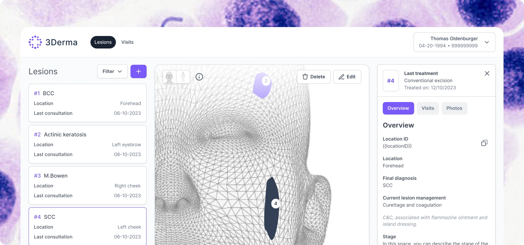
Put the 3D map at the center.
Every lesion sits as a numbered marker in its actual anatomical position, with a dedicated head view for the face, where most skin cancers appear. The list and the model stay in sync: click a card and the model highlights it, click a marker and the details open. Providers remember patients spatially, so the interface works the same way. Someone who has never seen this patient starts from the same picture as someone who has treated them for years.
Structured years of history, per lesion.
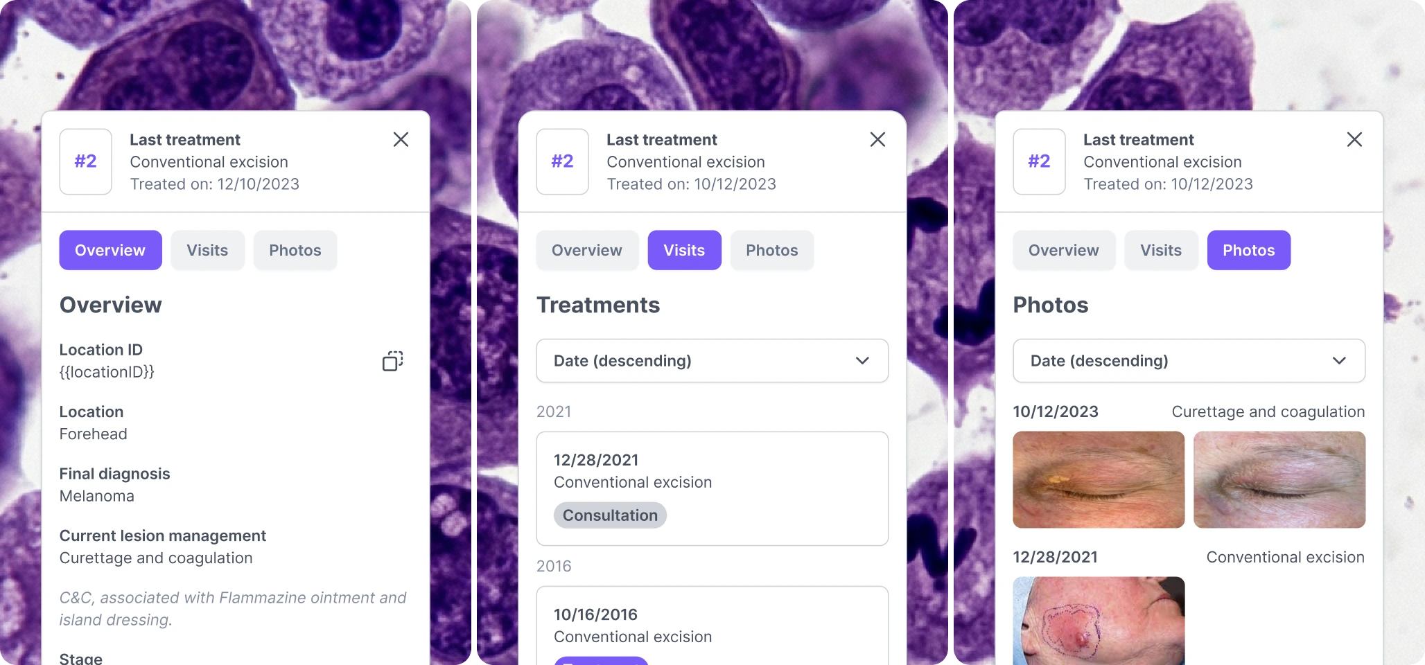
Each lesion opens into three views: an overview with diagnosis, treatment policy, dimensions and the latest report; a visit timeline; and a photo history. Visits are tagged as consult, treatment or result, so a provider can scan eight years of encounters and see what happened when, no matter who performed it. Photos from different years sit side by side, which is how you actually spot growth.
Built the record around real consults.
Treatment entries capture what the next provider will look up: procedure type, PA number, whether the excision was radical, and free-text reporting. The visit view links lesions treated in the same session, because that's how care happens. One appointment, several spots.
Designed the EHR handshake.
Dutch hospitals run on HIX, and 3Derma has to live alongside it, not replace it. Creating a lesion generates a location ID with a one-tap copy button to paste into HIX. A progress indicator tells the provider the sync takes about three minutes, so nobody sits there refreshing.
Kept the interface calm.
This runs during ten-minute consultations, often with the patient watching the screen. Restrained colour, generous type, one accent hue for lesion identity. The 3D model carries the visual weight and everything else stays out of the way.
The result
A patient's full skin cancer history: every lesion, every treatment, every photo, readable from one 3D model by every provider involved in their care. Built to fit the systems those providers already use.
Like this project
Posted Nov 3, 2025
kin cancer patients pass between providers for years. We designed 3Derma: every lesion pinned to a 3D body model, its full history one click deep.