The network for creativity
Join 1.25M professional creatives like you
Connect with clients, get discovered, and run your business 100% commission-free
Creatives on Contra have earned over $150M and we are just getting started
Back to feedPost
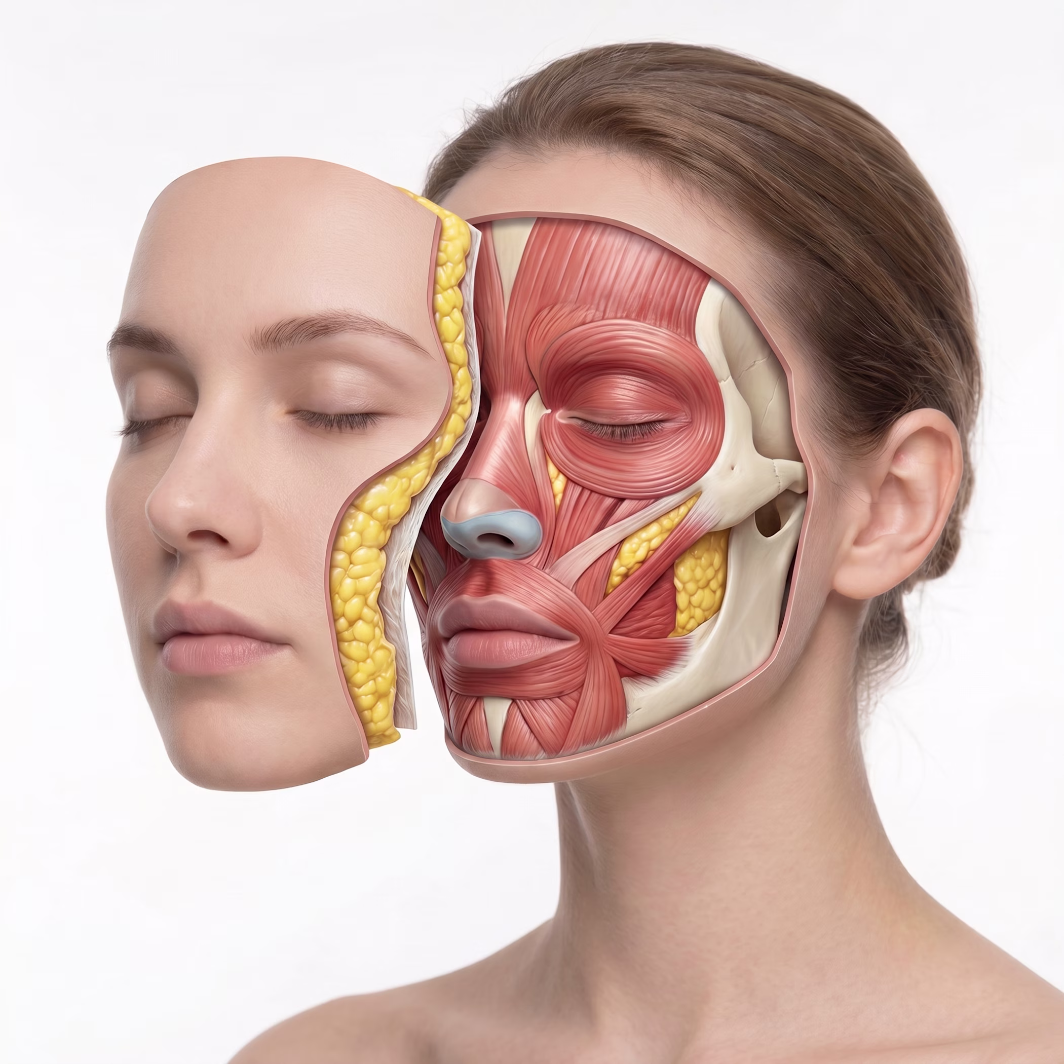
How Medical Illustrations Are Created
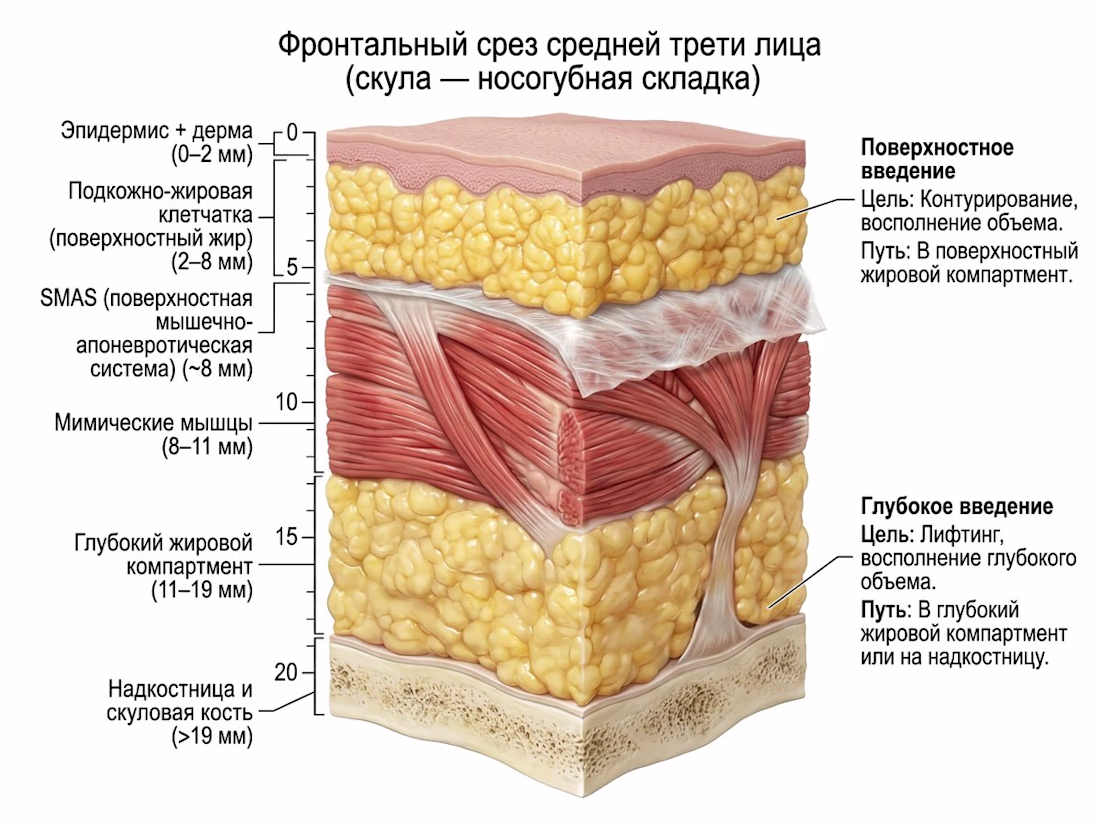
Every illustration begins not with a stylus, but with a stack of sources. Atlases, studies, surgical manuals. To illustrate facial tissue layers, this means checking every layer, every measurement, every structural relationship.
For a multilayered face, this means determining the location of the cross-section, how to show the different tissue types without reducing them to an unreadable diagram, how to use color to code depth. Then comes the structure, then the drawing, then a notation system that tells the surgeon not only what is there but also what it means for their needle.
The result works simultaneously in the lecture hall, in a surgical manual, and in a training course. Not decoration. A tool.
Available for medical atlas projects, clinical teaching content, and aesthetic medicine materials.
#medicalillustration #anatomicalart #facialanatomy #clinicalillustration #aestheticmedicine
The network for creativity
Join 1.25M professional creatives like you
Connect with clients, get discovered, and run your business 100% commission-free
Creatives on Contra have earned over $150M and we are just getting started
Related posts
As an illustrator, the biggest challenge is often a blank page. Endless possibilities, but never knowing where to start.
Today I took a slightly different approach to drawing. I gave myself twelve Post-it notes, three colours and one theme. The result was a playful set of nature-inspired illustrations with a quirky, psychedelic twist.
Sometimes unexpected work comes along when we set ourselves some constraints.
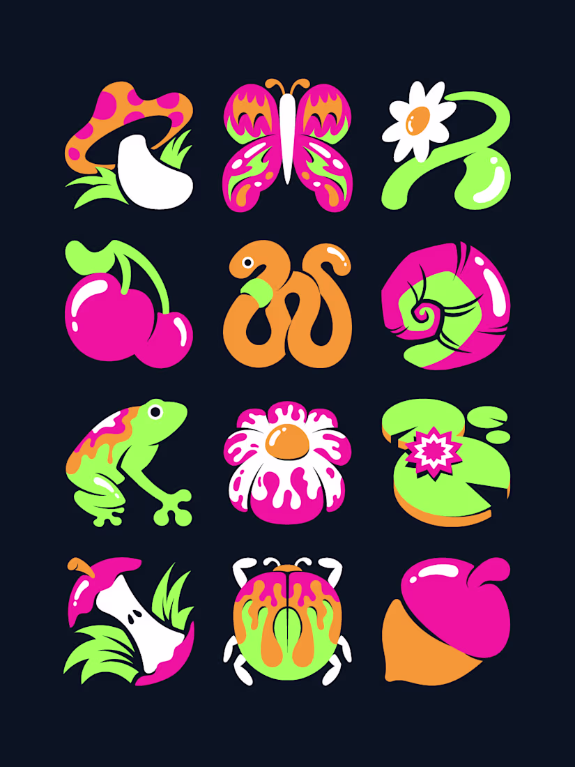
The mushroom snail hybrid is my favorite of the set, that shell pattern shouldn't work as well as it does. Constraints like this (twelve notes, three colors, one theme) seem to force way better ideas than a totally open brief. Do you do this kind of exercise regularly or was this a one off?
Good medical illustration doesn't dramatize. It clarifies.
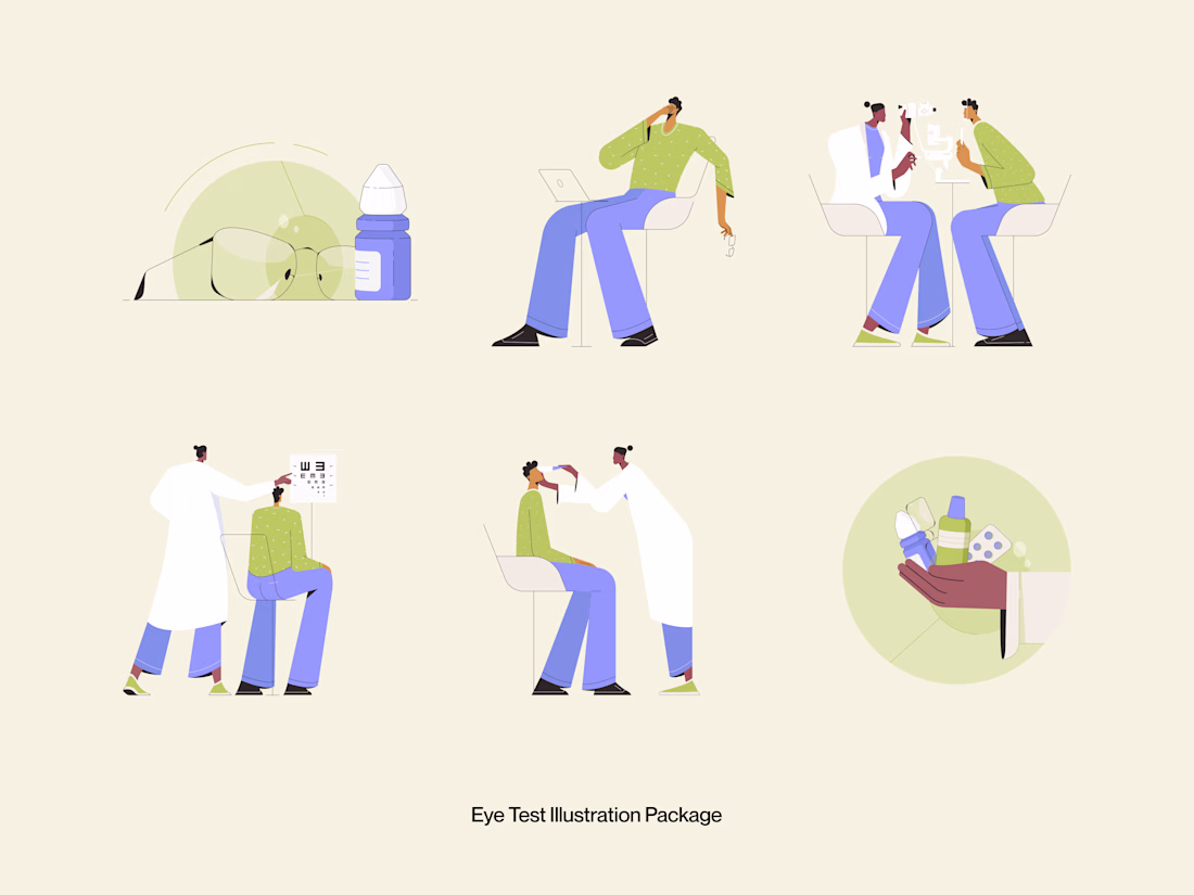
This Eye Test Illustration Package was built for healthcare platforms, optometry clinics, and health tech products that need warm, approachable visuals to communicate clinical moments without making them feel cold or intimidating.
Six scenes. Glasses and eye drops. A patient on a video call. Doctor-patient consultation. Vision chart test. Eye examination. Medication handoff. Every illustration covers a real touchpoint in the eye care journey, drawn in a style that feels human, not clinical.
Soft sage green, warm periwinkle blue, and a cream canvas that lets every character breathe. The color palette alone does half the work; it says "healthcare" without saying "hospital."
This is illustration work designed to live inside a product, not just decorate it.
Does this feel like a visual style your healthcare product needs? 👇
Tools: Illustrator · Figma
#IllustrationDesign #MedicalIllustration #FlatIllustration #HealthcareDesign #ContraFreelance #UIIllustration #DigitalIllustration #IconDesign
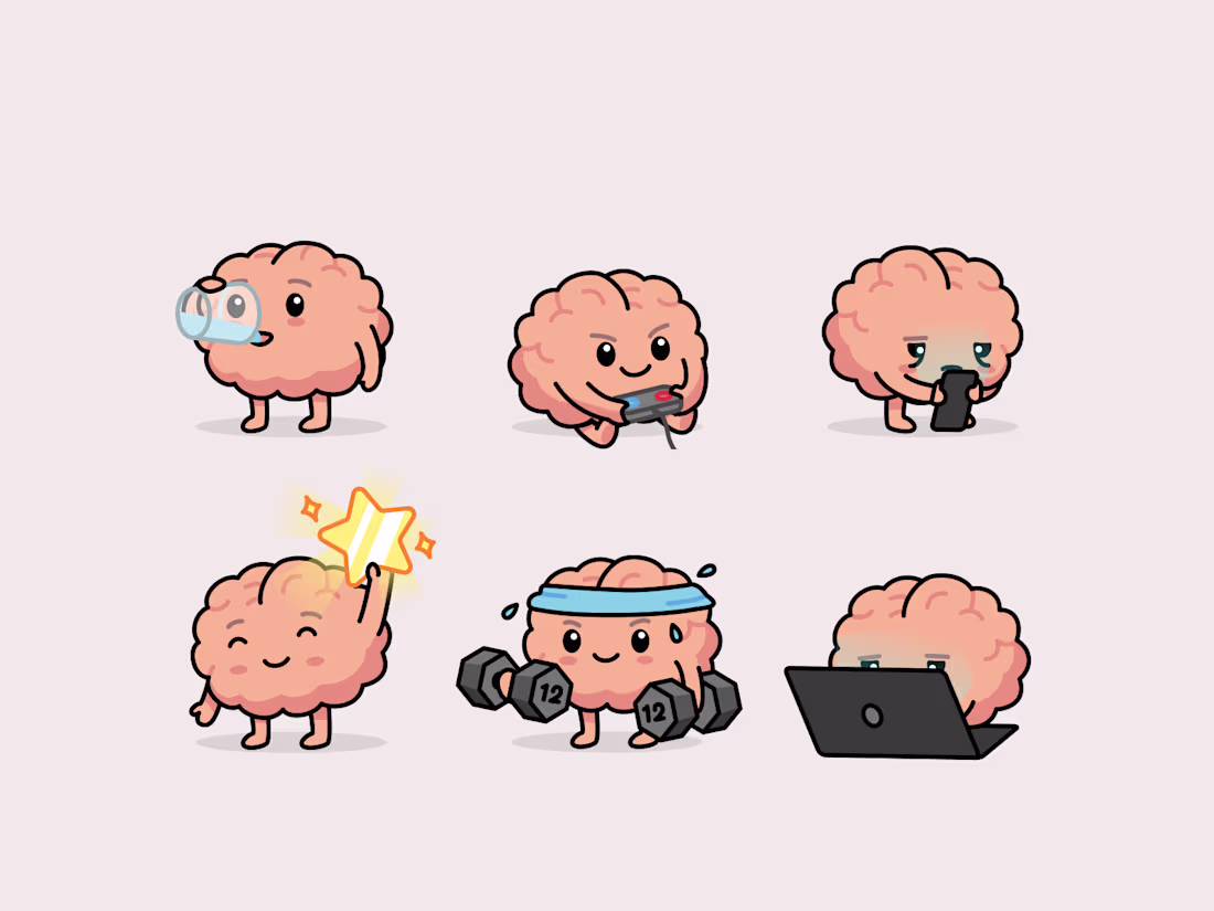
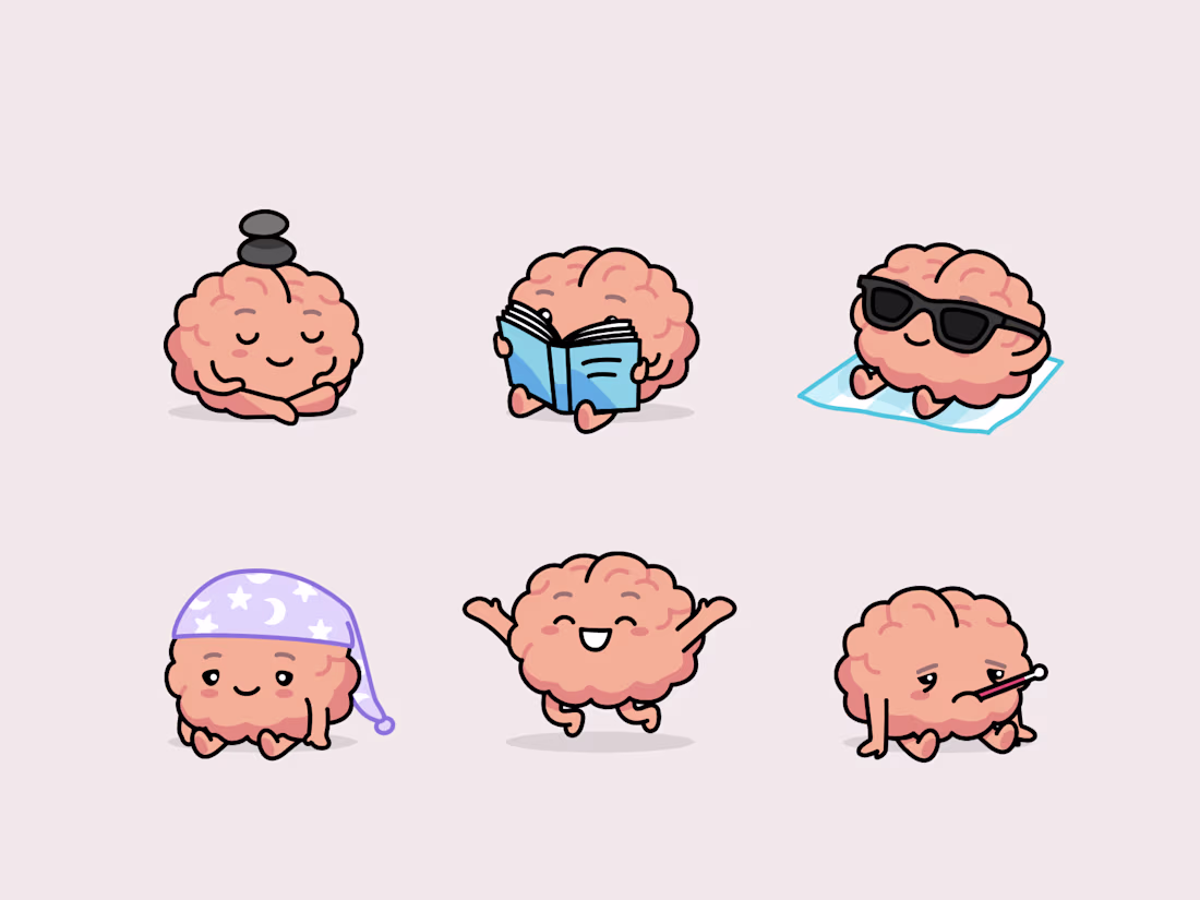
Nuggets everywhere!
We designed around 70 different Brains with @Mary Delaney
and then have @Arsen Airyan animate each and everyone of those!
If you brand or app needs a touch of cuteness, hit us up!
(mascotte brands lead to higher brand recognition, higher attention and ultimately higher sales! what are you waiting for?)

Absolutely love this!, The character designs are full of personality, and the animations bring them to life beautifully. It's inspiring to see such thoughtful branding and collaboration. Amazing work by the whole team! ❤️
Trending
Claude
Claude has entered the design space. How are you using Claude Design?
Contra University
Learn from expert creatives how to earn more using next-gen AI tools.
creativeaiflow
Creative AI workflows are evolving. What tools do you use, and what are their strengths and weaknesses?
freelancerlife
Freelancer life is wins, pivots, and everything in between. What’s yours right now?