
Albina Lavrenteva
Medical Designer-Illustrator
- $1k+
- Earned
- 2x
- Hired
- 5.00
- Rating
- 8
- Followers

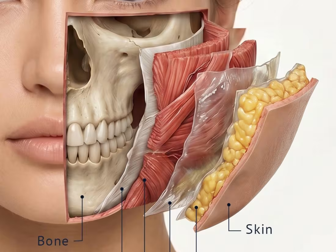
Six layers. One face. Everything a clinician needs to see — and rarely gets to.
Most people see a face as a surface. Every surgeon, injector, and aesthetic medicine practitioner works in the layers beneath it.
Bone. Periosteum. Muscle. SMAS. Superficial fat. Skin.
Each layer has its own mechanical properties, its own response to aging, its own role in how a face moves, holds its shape, and changes over time. Understanding the spatial relationship between these layers — not just their names — is what separates a technically precise practitioner from one who works by intuition.
This illustration and animation series visualizes all six facial layers in a single composition — peeling back from skin to bone, showing each layer in anatomical context.
Developed for aesthetic medicine education, plastic surgery training, and facial anatomy CME content.
If your organization creates training materials for cosmetic physicians, plastic surgeons, or aesthetic medicine educators — and needs medical illustration or animation that works at this level of anatomical detail — I'd love to discuss what's possible.
#medicalillustration (https://www.linkedin.com/search/results/all/?keywords=%23medicalillustration&origin=HASH_TAG_FROM_FEED) #medicalanimation (https://www.linkedin.com/search/results/all/?keywords=%23medicalanimation&origin=HASH_TAG_FROM_FEED) #facialanатomy (https://www.linkedin.com/search/results/all/?keywords=%23facialan%D0%B0%D1%82omy&origin=HASH_TAG_FROM_FEED) #SMAS (https://www.linkedin.com/search/results/all/?keywords=%23smas&origin=HASH_TAG_FROM_FEED) #aestheticmedicine (https://www.linkedin.com/search/results/all/?keywords=%23aestheticmedicine&origin=HASH_TAG_FROM_FEED) #plasticsurgery (https://www.linkedin.com/search/results/all/?keywords=%23plasticsurgery&origin=HASH_TAG_FROM_FEED) #facialaging (https://www.linkedin.com/search/results/all/?keywords=%23facialaging&origin=HASH_TAG_FROM_FEED) #CME (https://www.linkedin.com/search/results/all/?keywords=%23cme&origin=HASH_TAG_FROM_FEED) #medicaleducation (https://www.linkedin.com/search/results/all/?keywords=%23medicaleducation&origin=HASH_TAG_FROM_FEED) #medicalart (https://www.linkedin.com/search/results/all/?keywords=%23medicalart&origin=HASH_TAG_FROM_FEED)
0
88
Not every artery gets attention. This one deserves it.
The innominate artery — brachiocephalic trunk — is the first branch off the aortic arch. It feeds the right side of the brain and the right arm. When it fails, the surgical solution involves the arch itself.
This illustration visualizes prosthetic replacement of the innominate artery — graft anatomy, vessel relationships, surgical positioning.
Complex vascular anatomy needs precise visual support. Whether it’s for surgical training, CME content, or clinic education materials — that’s exactly what I do.
If you’re working on cardiology or vascular surgery education content and need an illustrator who understands the anatomy — reach out.
#medicalillustration (https://www.instagram.com/explore/tags/medicalillustration/) #vascularsurgery (https://www.instagram.com/explore/tags/vascularsurgery/) #aorticsurgery (https://www.instagram.com/explore/tags/aorticsurgery/) #cardiology (https://www.instagram.com/explore/tags/cardiology/) #innominateartery (https://www.instagram.com/explore/tags/innominateartery/)
1
100
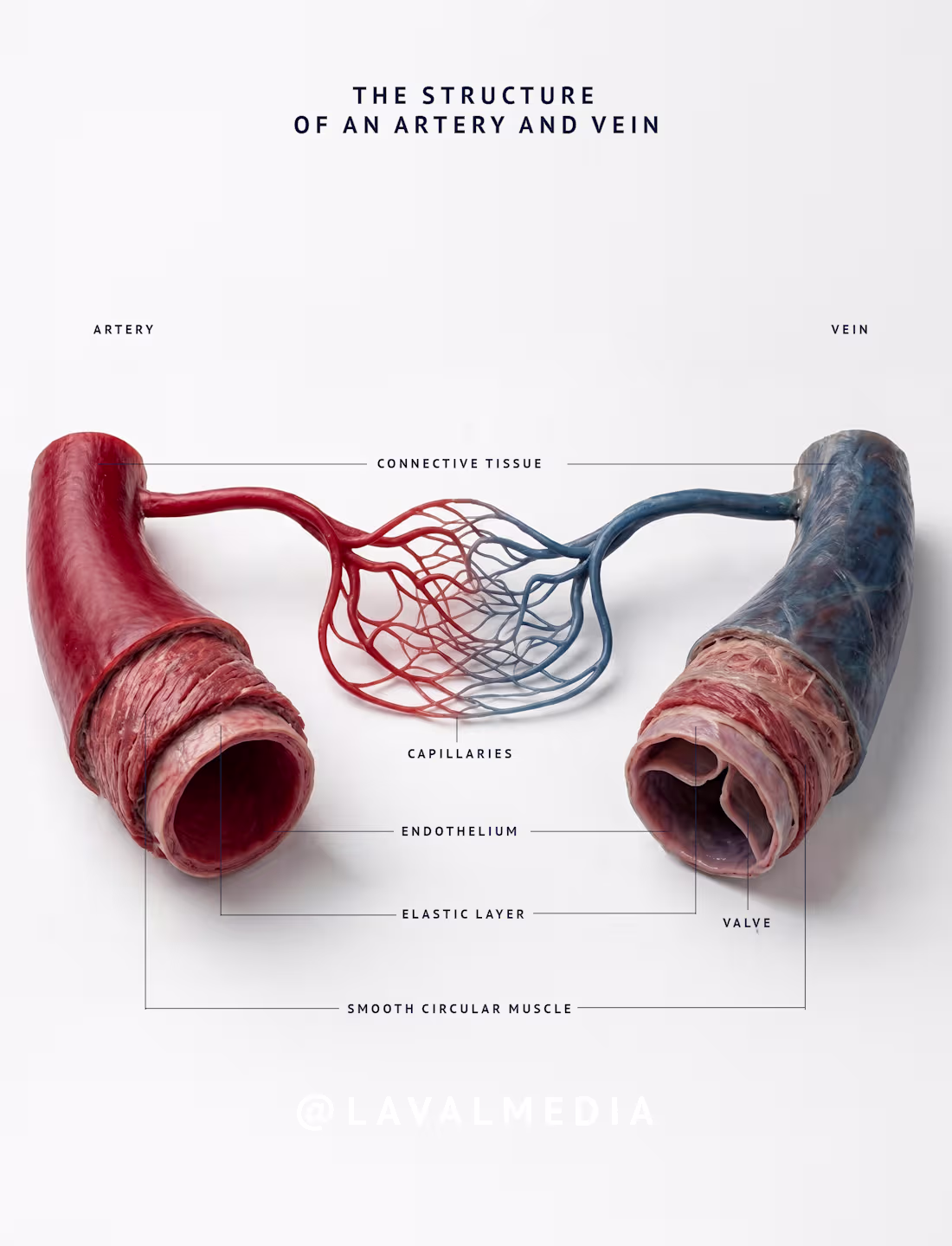
An artery. A vein. A capillary. Three completely different architectures.
The artery wall is built for pressure. Thick elastic layer, strong smooth muscle, connective tissue holding it all together. It expands with every heartbeat and springs back.
The vein carries the same blood, but at a fraction of the pressure. Thinner walls, less muscle, and something the artery doesn’t need: a valve. Because without it, blood would simply fall back down.
The capillary is just one cell thick. No muscle. No elastic layer. Just endothelium, because its entire job is exchange, not transport.
Same circulatory system. Completely different engineering solutions.
This illustration maps all three — layer by layer, structure by structure — the way it needs to be shown for medical education that actually teaches rather than just decorates.
If your team creates educational content for medical students, residents, or healthcare professionals — this is the level of detail that makes the difference.
#medicalillustration (https://www.instagram.com/explore/tags/medicalillustration/) #vasculararatomy (https://www.instagram.com/explore/tags/vasculararatomy/) #histology (https://www.instagram.com/explore/tags/histology/) #CME (https://www.instagram.com/explore/tags/cme/) #veinhealth (https://www.instagram.com/explore/tags/veinhealth/)
1
93
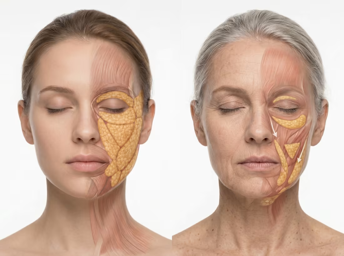
Look at these two faces.
Same person. Same anatomy. Just — time.
What changes isn't random. The fat pads that sit high on a young face don't disappear with age. They slide down. The ligaments that used to hold everything in place gradually let go. Volume shifts. Contours follow.
What you see on the surface — the fold, the shadow, the heaviness — is just the end of a story that started much deeper.
This illustration traces that process layer by layer — muscles, fat compartments, structural support — from a younger face to an older one.
For anyone working in aesthetic medicine or facial surgery — this is the part that actually matters.
#medicalillustration #facialaging #facialanatomy #aestheticmedicine #plasticsurgery #fatcompartments #medicalart
0
106
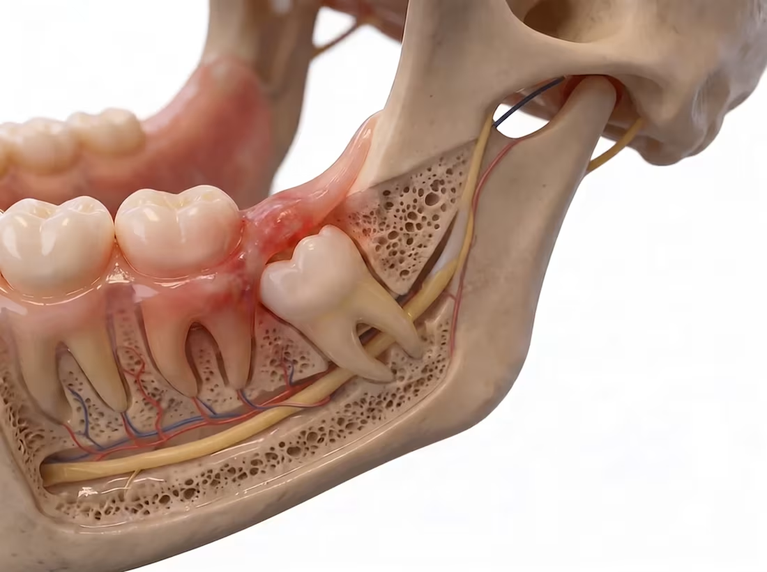
One tooth. One pressure point. One reason surgeons train on this anatomy.
The impacted third molar commonly called a wisdom tooth - is one of the most frequently illustrated subjects in oral surgery education. And for good reason.
When the third molar erupts at an angle and presses against the second molar, it creates a cascade of clinical consequences: pressure resorption of the adjacent root, periodontal pocket formation, increased caries risk, and in some cases - nerve involvement.
This illustration shows the mesial impaction in cross-section - the angulation, the contact point between the two roots, the surrounding bone, and the soft tissue relationship- everything a surgeon or dental student needs to understand before they enter the operatory.
That's what makes anatomically precise illustration valuable in dental education. Not the aesthetic. The clarity.
#medicalillustration #oralsurgery #dentalillustration #wisdomtooth #thirdmolar #dentalanatomy #CME #surgicaleducation #medicalart
0
92
Facial Vascular Anatomy — What Every Head & Neck Specialist Needs to Know
The face is one of the most vascularized territories in the human body. A dense network of arteries and veins — many of them communicating directly with the ophthalmic circulation — runs through every zone a surgeon, injector, or ENT specialist works in daily.
The facial artery crosses the mandibular border, ascends through the cheek, gives off labial branches, becomes the angular artery at the nasal ala, and communicates retrogradely with the ophthalmic artery. One vessel. Multiple disciplines. Multiple risk points.
Understanding this anatomy is not background knowledge. It is the difference between a safe procedure and a vascular complication.
This illustration shows the facial vascular system in surface projection — key arteries and veins mapped onto a realistic face, with foraminal exit points and zones of clinical risk clearly marked.
Part of a head & neck anatomical illustration series for clinicians, educators, and medical publishers working across maxillofacial surgery, aesthetic medicine, otolaryngology, and facial plastic surgery.
Precise anatomy. Built for clinical use.
Open to projects in medical illustration and anatomical visual systems.
#MedicalIllustration #FacialAnatomy #HeadAndNeck #VascularAnatomy #ClinicalIllustration
0
89
Three visual approaches to the same anatomical structure:
— Detailed anatomy — precise, anatomically accurate for studying structure
— Step-by-step schematic — focused on surgical stages and logical flow
— Graphic simplification — clean, minimal style for presentations and quick recall
Why this approach matters:
In medical education, visualization is not an add-on — it's essential. A student needs to do more than see; they need to understand structure, relationships, and surgical logic. Different learning styles require different visual approaches. One style cannot serve all educational goals.
Result:
The illustrations were integrated into the curriculum. Faculty reported that students became significantly more confident in navigating the anatomy of this complex region after working with the visual materials.
0
119
Complex anatomy made simple 👁️👃
Showing the invisible - that's our superpower!
Watch how animation and illustration can "uncover" the surface and take a peek inside the nasopharynx. From an artistic portrait to precise medical structures.
This video is a perfect example of how scientific illustration stops being boring and becomes art.
#animation #medicine #anatomicalart #nasopharynx #medicalillustration #aivideo #ai #anatomy
1
144
Animation demonstrating the structure of the head and the location of the arteries supplying the brain.
0
143

Demonstration of rhinoplasty surgery options. Image designed for a plastic surgeon.
1
149
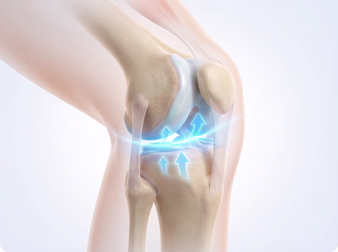
Medical infographics to demonstrate the benefits of a medical product
0
119
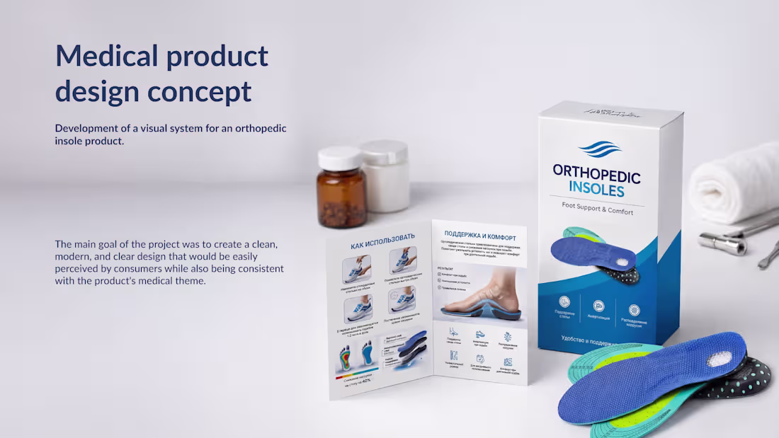
Development of a visual system for an orthopedic insole product. The project included the design of packaging, an information insert, and medical infographics explaining how the product works. The main goal of the project was to create a clean, modern, and clear design that would be easily perceived by consumers while also being consistent with the product's medical theme.
0
116
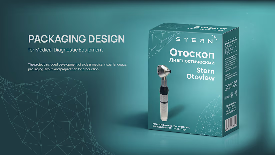
Packaging design for a diagnostic otoscope used in medical practice. The project included development of the visual concept, packaging layout and preparation of production-ready files. Two packaging dieline design concepts were proposed to explore different approaches to information hierarchy and product presentation
0
104