Embryonic-cardiac-Tissue-Profiling
Sabyasachi Chakrabarty
Project Overview
This project focuses on developing a computer vision solution using Deep Learning for the analysis of embryonic tissues. Enhanced tissue engineering workflows through computer vision approach. Provided efficient method for biologists to assess and improve tissue quality. The solution is implemented using PyTorch and consists of a two-phase model: Segmentation and Profiling.
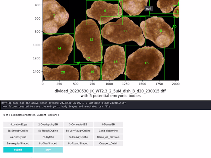
1. Segmentation Stage
Developed dynamic UNet Architecture from scratch: Designed to process high-resolution microscopy images, enabling precise segmentation of embryonic tissues.
Automated evaluation and quality enhancement of lab-cultivated embryonic tissues
Demonstrated AI's potential in medical image analysis
Integrated deep learning techniques in microscopy image processing
2. Profiling Phase
Developed custom CNN model for tissue property extraction from variable-resolution images
Its Built to accommodate variable image resolutions, allowing for the extraction of multiple tissue properties from segmented images.
Like this project
Posted Sep 25, 2024
Contribute to Sabya2/-Embryonic-cardiac-Tissue-Profiling development by creating an account on GitHub.
Likes
1
Views
14