OCT: The Intermarriage Between Imaging and Sampling.
·
4 min read
·
Mar 22, 2025

Healthy macula of a 24 year old male (cross-section view). Imaged in-vivo with an Optovue iVue Spectral Domain Optical Coherence Tomographer (SD-OCT) at the office of Drs. Harry Wiessner, Steven Davis, Daniel Wiessner, and Eric Wiessner in Walla Walla, WA, USA. 23 December 2013. This image is released to Wikimedia with patient consent. Photo adapted from ‘SD-OCT Macula Cross-Section’ by Wies6014, licensed under CC BY-SA 4.0 (https://creativecommons.org/licenses/by-sa/4.0/).
In medicine, plenty of imaging technologies exist to detect tissue pathology. One of the most advanced imaging technologies is OCT, or Optical Coherence Tomography. It has a high resolution that beats even MRIs. In fact, its tissue images are so precise and detailed that some have called it an “in-vivo sample,” in-vivo referring to “inside the body,” analogizing its resolution to that of a microscope. However, while OCT devices can reach resolutions close to those of optical microscopes, thanks to “infrared light interferometry,” it does come with limitations, namely its inability to penetrate deep into tissues, limiting its usefulness to imaging of superficial structures, such as the skin or the eyes.
Mechanism of OCT:
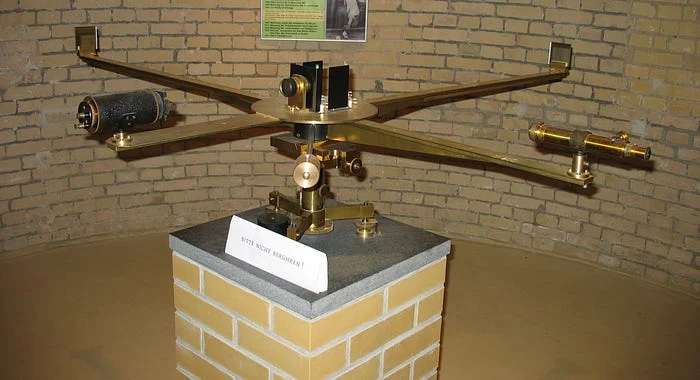
The basic idea of OCT is based on a historical device called Michelson’s Interferometer, where a source emits a coherent light beam that splits by means of an angled, half-transparent mirror into two light beams. One beam travels to the sample for an unknown distance (depending on the microscopic structure of the sample), then reflects back and is referred to as the sample beam. The other stream, the reference beam, travels a known distance to a reference mirror and reflects back. After these streams hit their respective targets and reflect, they combine, forming a unique “interference pattern” depending on the difference in the distance traveled by the two beams, which is dependent on the structure of the sample. Those patterns are then interpreted using a computer to construct a detailed image of the sample.

Michelson’s Interferometer: Historical reconstruction in the Michelson Cellar of the Michelson House on Telegraph Hill in Potsdam (the original setup no longer exists). The black cylindrical component on the left serves as the light source, emitting a beam that is split by a half-silvered mirror at the center. The two resulting beams travel along the perpendicular arms, reflect off mirrors at the ends, and then recombine to form an interference pattern. The golden tube on the right functions as an eyepiece or viewing telescope, allowing observers to analyze the resulting fringes. Photo adapted from ‘File:Michelsonnachbau.jpg,’ published on Wikimedia by Boson. Published under CC BY-SA 2.5 (https://creativecommons.org/licenses/by-sa/2.5/deed.en).
What does that Produce?
The core image produced by OCT is called an “A-Scan,” a one-dimensional image of the sample. Multiple A-scans along a specific plane of the sample are combined to produce a “B-Scan,” which is a two-dimensional cross-section image of the sample. Multiple B-scans can be combined to construct three-dimensional images of the sample, which comes in handy in assessing conditions where tissue atrophy or edema might be present.
Imaging speed is important in OCT devices because it allows for sharper images. It also enables the device to detect rapid changes in the sample, allowing for novel uses such as OCT Angiography, which assesses blood flow in the retina. This highlights the most significant differentiating factors between OCT types, imaging speed.
Types of OCT:
There are three main types of OCT:
Time Domain OCT: 1st gen. It uses a reference mirror that oscillates to gather depth information at various tissue layers. Thus, the imaging speed in this type is limited by the mechanical movement of the mirror.
Spectral Domain OCT: 2nd gen. It uses a spectrometer & Fourier transform algorithm to extract depth information at various layers simultaneously, enhancing its speed. It is the most commonly used type.
Swept Source OCT: 3rd gen. The light source rapidly sweeps across multiple wavelengths, also utilizing Fourier transform. It is the most advanced type in terms of speed and resolution.
The speed at which an OCT device can produce A-scans varies based on the type, ranging from 400 to 2,000 per second in Time Domain OCT and reaching up to 1,000,000 A-scans per second in Swept Source OCT.
Bridging the Gap between Microscopy and Imaging:
A useful way to think about OCT is that it’s the middle ground between conventional microscopes and traditional imaging methods like MRI or CT scans. Microscopes offer the best visualization of tissues, where a standard microscope can reach a resolution of 0.5 microns, meaning it can tell apart structures that are as close as half a micron. But the main problem is that it requires sampling, an invasive procedure where you take a tiny sliver of the tissue, process it, and then put it under the lens to examine its details. Because of this, a host of downfalls, like infections, lengthy sample preparations, sampling mistakes, the fact that you can only see a tiny sliver of the tissue, are associated with microscopic examination. On the other hand, traditional imaging methods can scan most of the body, they are quick, convenient, and instant. But they still fall short in terms of raw resolution. For example, while an average microscope can hit a resolution of 0.5 pretty easily, MRI, on the other hand, can only reach 1000 microns in terms of resolution. And this is where OCT comes in, it takes the high resolution advantage of microscopes, by utilizing infrared light interference, so much so that some advanced OCTs can now reach a resolution of 1 micron. It also takes the benefit of Imaging methods, like the speed, and the ease of use. However, OCT still falls short in terms of depth, compared to traditional methods. as it can only reach a depth of only around 1 cm, compared to other methods which can capture the whole body depth in one go. This is mainly because OCT uses infrared light, which doesn’t have the ability to penetrate deep into the tissues.
Conclusion:
In sum, OCT is a powerful imaging method that enables histology-level analysis of tissues without the need for invasive methods. However, it falls short of capturing deep structures due to the limitations of infrared light.
Need a walker, lift, braces, or wound care? CSA Medical Supplies has solid gear, shipped free in the continental U.S. 15 years of getting it done. Not happy? Return it within 60 days. Find a cheaper price within a week, and they’ll refund the difference. Check them out by clicking here. Keep in mind that this is an affiliate link, so we earn a commission if you buy using our link.
Like this project
0
Posted Mar 29, 2025
In medicine, plenty of imaging technologies exist to detect tissue pathology. One of the most advanced imaging technologies is OCT, or Optical Coherence Tomogr…
Likes
0
Views
0
Timeline
Mar 15, 2025 - Mar 20, 2025


