The network for creativity
Join 1.25M professional creatives like you
Connect with clients, get discovered, and run your business 100% commission-free
Creatives on Contra have earned over $150M and we are just getting started
Back to feedPost

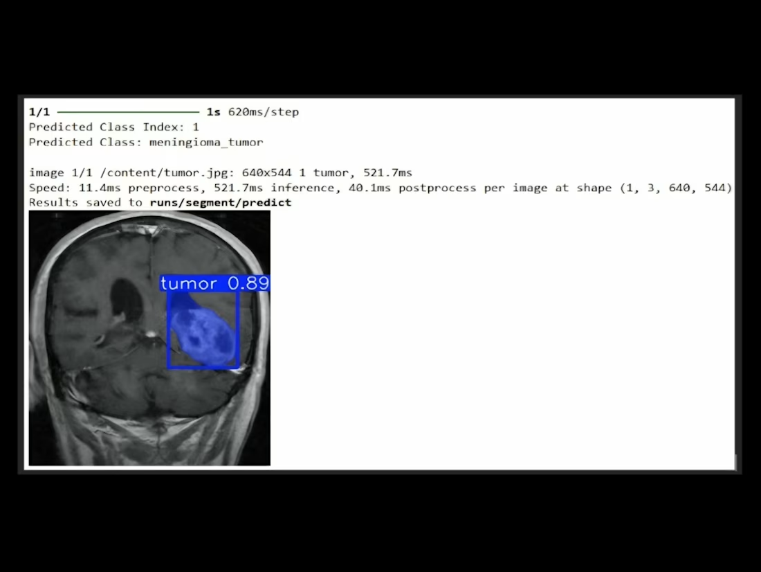
Brain Tumor Classification & Segmentation Model
Stage 01
Classification: A VGG16-based architecture, pretrained on ImageNet, was
fine-tuned with additional dense layers and dropout for tumor-type prediction. It also can easily represent spatial hierarchies of MRI images and
thus, is highly suitable in transfer learning due to its uniform architecture
with stacked convolutional layers. The last SoftMax layer delivers
four possibilities of classes.
Stage 02
Segmentation: The tumor regions were detected using a YOLOv8n instance
segmentation model trained on 10 epochs with an overlapping of the bounding box and class confidence scores.
Conditional
Workflow: A fundamental conditional logic is used to decide whether or not to perform segmentation.
The classification model uses the MRI images whereby the accurate region of interest (ROI) is extracted using a binary thresholding method to remove only the most useful features of analysis. The segmentation process will enable accurate tumor region segmentation, which plays a vital role in accurate diagnosis


The network for creativity
Join 1.25M professional creatives like you
Connect with clients, get discovered, and run your business 100% commission-free
Creatives on Contra have earned over $150M and we are just getting started




Related posts

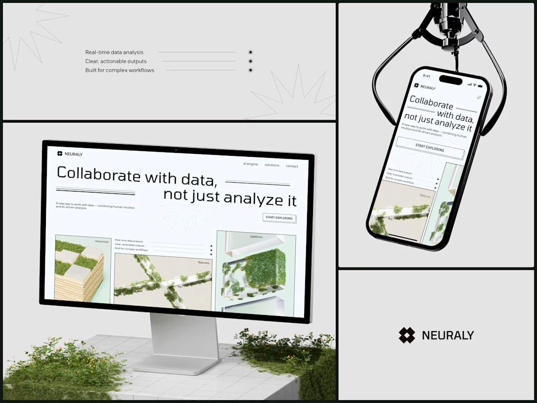
Neuraly - AI Data Analytics Platform
The headline says "Collaborate with data, not just analyze it" and the entire visual system is reinforcing that exact position. Data isn't cold infrastructure you feed queries into. It's something alive that grows when humans interact with it. The moss on architecture is literally that metaphor made visual: natural intelligence layered on top of built systems.
#AIDesign #WebDesign #B2BTech #BrandDesign #EditorialDesign


This one looks quite great honestly

Really nice concept and clean app design. The marketplace and community-focused experience look very thoughtfully put together. Great work! 🚀


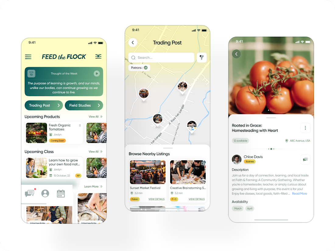
Feed The Flock — community platform for trading homegrown/homemade goods and sustainable-living education.

You Will Get Apollo + LinkedIn Sales Navigator B2B Leads, Verified Emails & Direct Phone Numbers.
Need targeted B2B leads with accurate contact information for your sales or outreach?
What You Provide:
✔Target industry / niche
✔Location
✔Company size or other filters
✔Decision-maker titles
✔Or your Apollo / LinkedIn Sales Navigator search link
What I Do:
I research and qualify the right companies and decision-makers, then enrich the prospects with:
✔Verified business emails
✔Direct phone / mobile numbers
✔LinkedIn profiles
✔Job titles
✔Company information
I also verify, clean, and remove duplicates before delivery.
What You Receive:
Company • Contact Name • Job Title • LinkedIn • Verified Email • Direct Phone • Website
Why Choose Me:
✓ Targeted B2B research
✓ Multiple data sources
✓ Manual data checking
✓ Email & contact verification
✓ Clean, organized, outreach-ready data
Tools: Apollo • LinkedIn Sales Navigator • Lusha • ContactOut • SalesQL • Google Sheets
Ready to Get Started?
Send me your target requirements or Apollo / Sales Navigator search link, and I can start with a small sample.


good work